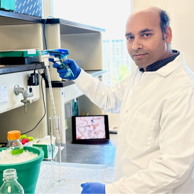
BMB Research Spotlight: Dr. Md Shafiqur Rahman
Dr. Md Shafiqur Rahman is a postdoctoral research associate in the laboratory of Professor Kelly Kim, and co-first author of the group’s latest publication appearing in the Journal of Molecular Biology: "Structural and Functional Characterization of Pseudomonas aeruginosa Virulence Factor AaaA, an Autotransporter with Arginine-Specific Aminopeptidase Activity."
The College of Natural Science spoke with Dr. Rahman to learn more about the team’s discoveries, as well the latest breakthroughs achieved by Spartan biochemists who are leveraging MSU's newly-expanded research facilities.
The Kim Lab studies membrane protein transport, and particularly how these crucial proteins arrive in the right place and time to support cellular function. Can you speak a bit more about the research happening in the lab and the questions you pursue?
Our lab is fascinated by how cells achieve spatial organization by localizing membrane proteins to appropriate organellar membranes—a process essential for normal cellular function. For membrane proteins to function properly, they not only need to be synthesized correctly, but also reach specific destinations (like organelles or the cell surface) to perform their roles in signaling, transport, or other critical processes.

Our lab investigates molecular mechanisms of how membrane proteins are targeted and inserted into appropriate membranes. We particularly focus on two systems: (1) the Guided entry of tail anchored protein (GET) pathway, a widely conserved pathway that mediates the targeting of tail-anchored proteins to the endoplasmic reticulum in eukaryotes, and (2) bacterial autotransporters, which are outer membrane proteins capable of self-translocation, many of which function as virulence factors in Gram-negative pathogens. By dissecting the mechanisms of these two systems, we aim to uncover universal principles of membrane protein biogenesis, with potential implications for understanding diseases and even drug targeting.
My current research focuses on elucidating how the insertase complex of the GET pathway, Get1/2, mediates the insertion of tail-anchored proteins into the ER membrane in Saccharomyces cerevisiae. In parallel, I investigate the structure and function of two bacterial autotransporters—AaaA from Pseudomonas aeruginosa and App from Neisseria meningitidis—to better understand their roles in protein translocation and pathogenesis.
What originally made you interested in this area of research?
My interest in bacterial autotransporters stems from my deep fascination with how
pathogens interact with their hosts at the structural and mechanistic levels—a theme
that has been central to my research journey. During my PhD, I worked extensively
on viral proteins, including the SARS-CoV-2 spike protein and HCV NS5A, using cryo-EM
and other structural biology techniques to uncover how these molecules facilitate
infection and can be targeted therapeutically.
What particularly excites me about autotransporters is how they represent an elegant
yet understudied example of bacterial virulence factors that share key conceptual
parallels with viral systems. Like the viral proteins I’ve studied, autotransporters
are modular, with distinct domains mediating functions like membrane translocation
(the β-barrel) and host interaction (the passenger domain). However, their autonomous
secretion mechanism adds an additional layer of complexity that I find intellectually
compelling.
My experience in determining high-resolution structures of challenging targets—such
as membrane-associated viral proteins and aptamer-protein complexes—has equipped me
with the technical skills to investigate autotransporters. Moreover, my background
in structure-guided therapeutic development (e.g., designing bivalent aptamers against
SARS-CoV-2) makes me eager to explore how autotransporter structures could inform
new anti-virulence strategies.
Ultimately, I see autotransporters as a perfect bridge between my past work and future
goals: they combine my expertise in membrane protein biology, structural analysis,
and translational applications, while offering new mechanistic puzzles to solve. I’m
particularly motivated to contribute to understanding how these systems evolve to
mediate host-pathogen interactions—and how we might disrupt them.
Your latest publication sheds light on a membrane protein found in Pseudomonas aeruginosa — a bacterium that can cause a range of diseases in humans, plants, and animals. What exactly did the team discover?
We determined the first high-resolution cryo-EM structure of the autotransporter AaaA from Pseudomonas aeruginosa and demonstrated that it is a highly specific, zinc-dependent aminopeptidase that cleaves only N-terminal arginine from peptides. Our structure revealed that AaaA differs from conventional autotransporters, featuring an uncommon architecture—a compact globular domain responsible for aminopeptidase attached to a β-barrel membrane anchor. Mutational analysis identified key residues critical for binding and cleaving N-terminal arginine, while biochemical assays showed that AaaA retains robust activity across a wide range of pH and temperatures, and relies strictly on zinc. These findings clarify how AaaA enables efficient arginine utilization in P. aeruginosa, supporting bacterial survival and virulence, and highlight its potential as a target for antimicrobial therapies.
What do you find most exciting about these findings?

What’s most exciting about these findings is that we’ve uncovered the molecular basis
for exactly how the autotransporter AaaA from P. aeruginosa achieves its unique, highly
specific enzymatic activity. We discovered that AaaA is not only a robust, surface-displayed
enzyme, but also the only structurally unique autotransporter with a compact globular
fold and strict selectivity for N-terminal arginine. By resolving its high-resolution
structure and pinpointing the key residues and zinc ions that determine this specificity,
we revealed a precise mechanism by which P. aeruginosa uses AaaA to extract arginine
as a nutrient—directly linking its enzymatic features to bacterial survival and virulence.
This molecular insight not only broadens our fundamental understanding of how bacterial
pathogens scavenge nutrients but also opens a clear path for developing targeted inhibitors
that could weaken infections by blocking AaaA’s activity. In short, we’ve provided
a blueprint for both new science and tangible therapeutic innovation.
AaaA is a surface-exposed autotransporter in the outer membrane of Pseudomonas aeruginosa,
with a compact globular passenger domain that has a zinc-dependent active site that
specifically cleaves N-terminal arginine residues from peptides and the C-terminal
β-barrel translocator domain embedded in the outer membrane, according to a graphical
abstract from the Kim Lab's most recent paper in the Journal of Molecular Biology.
How do you see these discoveries contributing to larger conversations related to organism health and future applications?
These discoveries advance our understanding of how P. aeruginosa acquires arginine to support survival and virulence during infection, highlighting AaaA’s unique structure and zinc-dependent specificity as key to this process. This insight into nutrient scavenging enriches broader discussions on pathogen-host interactions and metabolic adaptation in bacterial pathogenesis. Moreover, identifying AaaA as a surface-exposed, structurally distinct enzyme with essential catalytic residues opens new avenues for designing targeted antimicrobials that disrupt nutrient uptake, offering promising strategies to combat infections while potentially minimizing antibiotic resistance.
The Kim Lab leverages cryo-electron microscopy to achieve its science. What makes these tools so useful or powerful for researchers, especially knowing MSU’s own cryo-EM facility is just now completing a substantial expansion?
Cryo-electron microscopy (cryo-EM) has been transformative for structural biology because it lets us visualize intricate molecular machines—like membrane protein targeting complexes or bacterial autotransporters—in near-native states at atomic resolution. Unlike traditional methods that often require crystallization, cryo-EM captures snapshots of proteins frozen mid-action, revealing dynamic conformations essential for their function. It is particularly powerful for studying large, flexible, or membrane-associated proteins that are challenging for X-ray crystallography or NMR, and it can do so with relatively small amounts of sample while accommodating heterogeneous or transient complexes.
With MSU’s cryo-EM facility expanding its high-end instrumentation, we can now determine protein structures more efficiently and tackle increasingly ambitious questions. This upgrade positions MSU researchers to lead in structural biology, combining speed, resolution, and the ability to study “uncooperative” proteins that resist other techniques. For example, our lab plans to use Titan Krios, the new cryo-EM microscope in the facility, to determine the structure of other autotransporter proteins like App of N. meningitidis, and H. pylori’s FaaA to understand their roles in pathogenesis.
Where might this research take you next? Are there unanswered questions the Kim Lab would like to answer?
Building on our structural work with the AaaA autotransporter, now I am investigating the App autotransporter from Neisseria meningitidis, a key virulence factor in bacterial meningitis. Using structural and biochemical approaches, we aim to reveal how App's unique passenger domain mediates pathogenesis, with particular focus on its protein translocation mechanism and potentially exposing new drug targets.
In parallel, I am also studying the eukaryotic Get1/2 insertase complex to understand its role in directing tail-anchored proteins to the ER membrane. This work provides an evolutionary counterpoint to bacterial autotransporters, revealing how different organisms solve the universal challenge of membrane protein biogenesis.
Through comparative study of bacterial autotransporters and eukaryotic Get1/2 insertase, we aim to uncover universal principles of membrane protein biogenesis, understand diseases and develop targeted antimicrobial strategies that disrupt pathogen virulence mechanisms.


